One-Pager
Shock Types at a Glance
Hemodynamic patterns, exam findings and first treatments for each shock type
Download one-page PDFLetter landscape, ready to print or keep with your notes
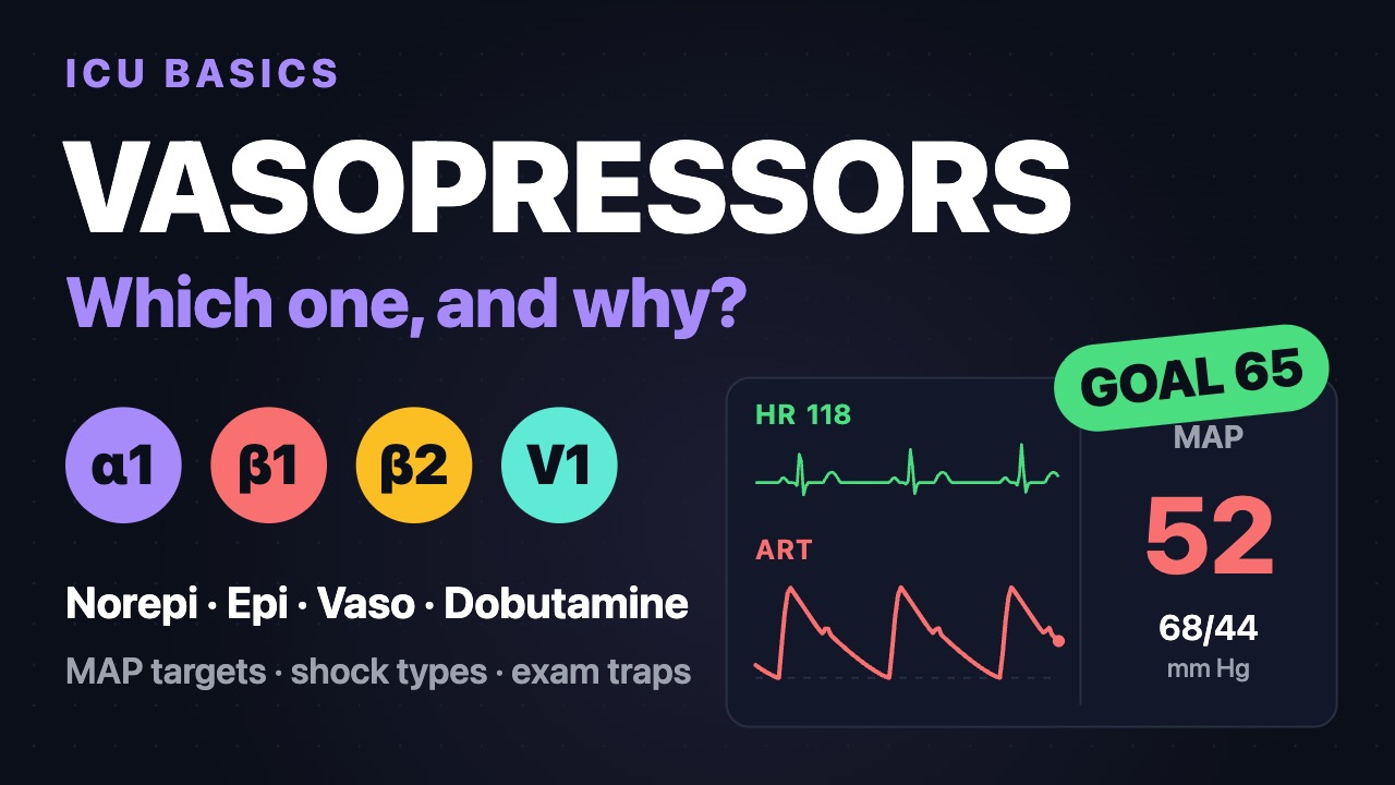
Watch the explainer
Hemodynamic patterns
Shock: cells get or use too little oxygen; hypotension is not required. Hypovolemic, cardiogenic and obstructive shock are low-flow; distributive is low-resistance. Lactate >2 mmol/L marks hypoperfusion in any type; trend it to judge response.
| Type | CO / SVR | Wedge / CVP | SvO2 |
|---|---|---|---|
| Hypovolemic | CO low, SVR high | Both low | Low |
| Cardiogenic (LV) | CO low, SVR high | Both high | Low |
| Obstructive: PE | CO low, SVR high | CVP high, wedge low or normal | Low |
| Obstructive: tamponade | CO low, SVR high | Both high, nearly equal | Low |
| Septic | CO normal or high, SVR low | Low or normal | Normal or high |
| Anaphylactic or neurogenic | CO low or normal, SVR low | Low | Normal or low |
Findings and first treatments
| Type | Exam findings | First treatments |
|---|---|---|
| Hypovolemic | Cool, pale, clammy; flat neck veins; narrow pulse pressure | Stop the loss; 1:1:1 blood products for bleeding; crystalloid otherwise |
| Cardiogenic | Cool, mottled; crackles, JVD; low urine output | Reperfuse an MI; norepinephrine; add an inotrope; MCS if failing |
| Obstructive | JVD plus muffled heart tones, absent breath sounds or sudden hypoxemia | Relieve it: chest decompression, pericardiocentesis, reperfusion for massive PE |
| Septic | Warm, flushed early; fever; cold late | Antibiotics within 1 h; ≥30 mL/kg balanced crystalloid in 3 h; norepinephrine |
| Anaphylactic | Hives, angioedema, wheeze or stridor | IM epinephrine first; IV fluids; epinephrine infusion if refractory |
| Neurogenic | Warm, dry skin; slow heart rate; cord injury T6 or above | Fluids, then norepinephrine; atropine for symptomatic bradycardia |
Septic shock (Sepsis-3): vasopressors needed for MAP ≥65 mm Hg and lactate >2 mmol/L despite adequate fluids. SSC 2026 MAP goal: 65 (60–65 at age 65 or older).
SCAI SHOCK stages (cardiogenic)
| Stage | Meaning | Typical findings |
|---|---|---|
| A: At risk | AMI or heart failure, perfusing well | Warm, normal lactate, SBP ≥100 (or baseline) |
| B: Beginning | Unstable, still perfusing | SBP <90, MAP <60 or >30 drop from baseline; HR ≥100; normal lactate |
| C: Classic | Hypoperfused; needs a drug or device | Lactate ≥2, cold, urine <30 mL/h; CI <2.2, PCWP >15 |
| D: Deteriorating | Failing initial support | Rising lactate; more pressors or MCS added |
| E: Extremis | Circulatory collapse | CPR or near pulseless; lactate ≥8, pH <7.2 |
Cardiac arrest modifier: add only if comatose (GCS <9) after the arrest. Current US guidance: norepinephrine first; no routine IABP in MI-related shock.
Neurogenic vs spinal shock
| Feature | Neurogenic shock | Spinal shock |
|---|---|---|
| What is lost | Sympathetic tone | Reflexes and function below the injury |
| Findings | Low BP, low HR, warm skin | Flaccid paralysis, areflexia, no sensation |
| Shock? | Yes, distributive | No, not circulatory |
| Duration | Often 48–72 h, can last weeks | About 4–6 weeks; ends when the bulbocavernosus reflex returns |
Cord injury MAP: a 2024 guideline suggests at least 75–80, not above 90–95, for 3–7 days. Many exam items still use 85–90 for 7 days.
Reading the numbers on exams
- Low SVR: distributive. Slow HR suggests neurogenic; hives or wheeze, anaphylaxis; infection, sepsis.
- High SVR, low CO: low-flow; check the filling pressures.
- CVP and wedge low: hypovolemic. Wedge above about 15–18: cardiogenic.
- High CVP with a low or normal wedge: PE or RV infarct.
- CVP, PA diastolic and wedge high and nearly equal: tamponade, with Beck triad and pulsus paradoxus (inspiratory SBP fall >10 mm Hg).
In depth
The one-page summary above is what the PDF prints. These sections go further.
Oxygen delivery, consumption and lactate
Every shock type ends in the same place: oxygen delivery (DO₂) falls below what the tissues consume (VO₂), or the cells cannot use what arrives. CaO₂ = (1.34 × Hb × SaO₂) + (0.003 × PaO₂), about 20 mL/dL. DO₂ = CO × CaO₂ × 10, about 1,000 mL/min (DO₂I 500–600 mL/min/m²). Resting VO₂ is about 250 mL/min, so the oxygen extraction ratio (O₂ER = VO₂ ÷ DO₂) is about 25% and SvO₂ sits near 70–75%.
Worked example: Hb 15, SaO₂ 98% and CO 5 L/min give DO₂ 1,000 mL/min. After hemorrhage to Hb 7 with SaO₂ 90% and CO 3, CaO₂ is 8.6 mL/dL and DO₂ about 260 mL/min, a 74% fall. Raising PaO₂ from 100 to 300 adds only 0.6 mL/dL; hemoglobin, saturation and flow are the levers.
As DO₂ falls, extraction rises and VO₂ holds steady while SvO₂ drops. Extraction tops out near 60–70%; below that point, the critical DO₂, VO₂ becomes supply-dependent and lactate rises. Critical DO₂ was about 330 mL/min/m² in anesthetized adults (Shibutani 1983) and about 4 mL/kg/min in dying ICU patients (Ronco 1993); it varies and cannot be measured at the bedside, so lactate and SvO₂ trends stand in for it.
A low SvO₂ means low CO, Hb or SaO₂, or high consumption (fever, shivering, work of breathing). A high SvO₂ in shock means the cells are not extracting: sepsis with microcirculatory shunting, cyanide or carbon monoxide, or a wedged catheter. ScvO₂ normally reads 2–3% below SvO₂ but 5–8% above it in shock, so 70% can hide an SvO₂ in the low 60s. A venous-to-arterial PCO₂ gap above 6 mm Hg flags inadequate flow even with a normal ScvO₂ (Cecconi 2014).
| Lactate type | Mechanism | Examples |
|---|---|---|
| Type A | Tissue hypoxia or hypoperfusion | Any shock, severe anemia, CO poisoning, seizures |
| Type A (regional) | Local ischemia | Mesenteric ischemia, compartment syndrome |
| Type B1 | Underlying disease | Liver failure, malignancy, thiamine deficiency |
| Type B2 | Drugs and toxins | Epinephrine, albuterol, metformin, propofol, cyanide |
| Type B3 | Inborn errors of metabolism | Mitochondrial and glycogen storage disorders |
The liver and kidneys clear lactate, so liver failure keeps it high after perfusion is restored. Epinephrine raises lactate through β₂-driven aerobic glycolysis; a rising lactate on epinephrine with improving perfusion is usually type B. Sepsis-3 uses >2 mmol/L; SSC 2021 used ≥4 mmol/L, or hypotension, to trigger the 30 mL/kg bolus. Repeat within 2–4 h and aim for a 10–20% fall every 2 h.
Hypovolemic and hemorrhagic shock
Lost volume lowers venous return and stroke volume; baroreceptors raise heart rate and SVR. Vasoconstriction holds diastolic pressure while systolic falls, so pulse pressure narrows before SBP drops, and young adults can lose 30% of blood volume first. Tachycardia, cool skin and a shock index (HR ÷ SBP) above 0.9–1.0 are the early signs. Non-hemorrhagic losses (GI, burns, DKA, diuresis) get balanced crystalloid (SMART 2018) and treatment of the cause.
| Class | Blood loss | HR / SBP / pulse pressure | Urine, mental status, base deficit |
|---|---|---|---|
| I | <15% (<750 mL) | <100 / normal / normal | >30 mL/h; slightly anxious; 0 to −2 |
| II | 15–30% (750–1,500 mL) | 100–120 / normal / narrowed | 20–30 mL/h; anxious; −2 to −6 |
| III | 30–40% (1,500–2,000 mL) | 120–140 / low / narrowed | 5–15 mL/h; confused; −6 to −10 |
| IV | >40% (>2,000 mL) | >140 / very low / narrowed | Negligible; lethargic; −10 or worse |
- Stop the bleeding: pressure, tourniquet, pelvic binder, then OR or interventional radiology.
- Activate massive transfusion early: ABC score ≥2 (penetrating injury, SBP ≤90, HR ≥120, positive FAST) or shock index >1.
- Plasma, platelets and red cells 1:1:1, or low-titer O whole blood; minimal crystalloid.
- Tranexamic acid 1 g over 10 min within 3 h of injury, then 1 g over 8 h (CRASH-2); not started after 3 h.
- Permissive hypotension (SBP 80–90) until control, except with brain injury: Brain Trauma Foundation 2016 targets SBP ≥100 at age 50–69 and ≥110 otherwise.
- Keep temperature >36 °C and ionized calcium >1.1 mmol/L (citrate binds it), correct acidosis, and guide products with TEG or ROTEM.
Cardiogenic shock: LV, RV, SCAI and MCS escalation
Cardiogenic shock is hypoperfusion from pump failure: classically SBP <90 for ≥30 min or pressors needed, CI <2.2 L/min/m² and PCWP >15. A rising LVEDP backs up into the lungs and lowers coronary perfusion pressure (aortic diastolic minus LVEDP), worsening ischemia. In current US registries (Critical Care Cardiology Trials Network) heart-failure shock outnumbers AMI shock.
Cardiac power output, CPO = MAP × CO ÷ 451, is normally about 1 W; below about 0.6 W predicts death (Fincke 2004). PAPi = (PA systolic − PA diastolic) ÷ RAP flags RV failure below 0.9 in inferior MI (Korabathina 2012). SCAI stages are re-scored as the patient changes; in-hospital mortality rose from about 3% at stage A to about 67% at E in a cardiac ICU cohort (Jentzer 2019). Moving from C to D is the usual shock-team trigger for mechanical support.
RV shock follows RV infarct (inferior STEMI, ST elevation in V4R), PE, ARDS, pulmonary hypertension and LVAD implantation. The dilated RV pushes the septum into the LV, so LV filling falls even with normal LV muscle. Give fluid until CVP is about 10–15 mm Hg, then stop; keep AV synchrony (pace heart block); avoid hypoxemia, hypercapnia, acidosis and high PEEP, which raise PVR; add dobutamine or milrinone and inhaled pulmonary vasodilators; escalate to Impella RP, ProtekDuo or VA-ECMO.
- Reperfuse AMI shock at once, culprit lesion only (CULPRIT-SHOCK).
- Norepinephrine first (SOAP II); avoid dopamine and use epinephrine cautiously (OptimaCC: refractory shock 37% vs 7% with norepinephrine).
- Add an inotrope once MAP holds: dobutamine 2.5–10 mcg/kg/min or milrinone 0.125–0.5 mcg/kg/min (renally cleared, vasodilates; DOREMI 2021 found no difference).
- Diurese or start CRRT for congestion; expect hypotension at intubation.
- Use a PA catheter when type or response is unclear.
- Escalate to MCS at SCAI D, CPO <0.6 W or rising lactate on two agents, with a stated goal: recovery, durable VAD or transplant, or decision.
| Device | Support | Effect on the heart | Nursing focus |
|---|---|---|---|
| IABP | About 0.5–1 L/min | Lowers afterload, raises diastolic pressure | Timing, limb checks, platelets |
| Impella CP | About 3.5–4.3 L/min | Unloads the LV directly | Position, purge, hemolysis, limb |
| Impella 5.5 | Up to about 5.5–6 L/min | Full LV unloading; axillary access | Position, purge; can mobilize |
| Impella RP / ProtekDuo | About 4 L/min right side | Bypasses the failing RV to the PA | Hemolysis, position, RV filling |
| Peripheral VA-ECMO | 4–6+ L/min | Full flow but raises LV afterload | Limb, bleeding, LV distension |
Peripheral VA-ECMO returns blood up the aorta, so a stunned LV may not open the aortic valve: pulse pressure narrows below about 10 mm Hg, PCWP rises and pulmonary edema follows. An Impella (ECPELLA) or IABP vents it. Current US guidance (2025 ACC/AHA ACS guideline, as summarized in the ACC 2026 Year in Review) rates routine IABP Class 3 and the microaxial pump Class 2a for selected STEMI shock.
Obstructive shock: PE, tamponade, tension pneumothorax
Obstructive shock is a mechanical block to filling or ejection: pressures upstream are high, CO is low, and only relieving the obstruction fixes it.
Massive PE: a sudden rise in PVR dilates the thin RV, which cannot generate a mean PA pressure much above 40 mm Hg acutely; the septum bows into the LV and RV ischemia spirals into arrest. ESC 2019 high-risk PE is SBP <90 for ≥15 min, pressor need or arrest. Echo shows RV:LV >1, a D-shaped septum and McConnell sign. Give alteplase 100 mg over 2 h (50 mg bolus in arrest), or catheter or surgical embolectomy if lysis is contraindicated (ESC 2019; AHA/ACC 2026 rates all three 2a with persistent hypotension); VA-ECMO bridges. Use norepinephrine, limit fluid to about 500 mL, and avoid intubation if possible: induction and positive pressure can tip the RV into arrest.
Tamponade: pericardial pressure above chamber diastolic pressure stops filling. The right atrium collapses first; RV diastolic collapse is more specific. CVP, PA diastolic and PCWP equalize within about 5 mm Hg and pulsus paradoxus exceeds 10 mm Hg. A rapid 100–200 mL can cause it; a slow effusion can reach liters. Treat with echo-guided pericardiocentesis or surgical drainage, volume as a bridge, and no positive pressure if avoidable.
After cardiac surgery, tamponade is often a localized clot that echo can miss: chest tube output stops or slows while CVP rises and CO, urine output and MAP fall. The treatment is re-exploration, often at the bedside.
Tension pneumothorax: trapped air shifts the mediastinum and stops venous return. On the ventilator, peak and plateau pressures rise and SpO₂ falls before hypotension; tracheal deviation is late. Decompress without waiting for imaging: ATLS 10th edition uses the 4th–5th intercostal space just anterior to the midaxillary line in adults, and many EMS protocols still use the 2nd intercostal space midclavicular. A chest tube follows.
Distributive shock: septic, anaphylactic, neurogenic, adrenal
Septic shock combines vasodilation (inducible nitric oxide, relative vasopressin deficiency), capillary leak, microcirculatory shunting and mitochondrial failure; septic cardiomyopathy can turn warm shock cold. The sequence: cultures and antimicrobials within 1 h; balanced crystalloid with reassessment (passive leg raise raising CO ≥10%, or pulse pressure variation >12–13% in a passive, sinus-rhythm patient on VT ≥8 mL/kg); norepinephrine to MAP 65 (peripheral access is acceptable at first); vasopressin 0.03 units/min second (SSC 2021 suggested adding it at norepinephrine 0.25–0.5 mcg/kg/min); hydrocortisone 200 mg/day for an ongoing pressor need; epinephrine for a failing heart; source control.
Anaphylaxis: mast cell mediators cause vasodilation, rapid capillary leak and bronchospasm. IM epinephrine comes first, with 1–2 L of rapid crystalloid and the patient supine with legs raised; sudden sitting or standing can precipitate arrest. Shock persisting after two or three IM doses needs an epinephrine infusion, starting around 0.05–0.1 mcg/kg/min. Patients on beta blockers may need glucagon 1–5 mg IV over 5 min, then 5–15 mcg/min.
Neurogenic shock: injury above about T6 cuts sympathetic outflow (T1–L2), and loss of the cardiac accelerator fibers (T1–T4) leaves unopposed vagal tone, so suctioning or turning can trigger severe bradycardia or asystole. In trauma, hypotension is hemorrhage until proven otherwise. Give fluid, then norepinephrine, atropine 1 mg every 3–5 min to 3 mg for symptomatic bradycardia (AHA 2020), and pacing if refractory. Patients lose thermoregulation below the lesion.
Adrenal crisis: without cortisol, vessels stop responding to catecholamines, so shock resists fluids and pressors. Clues are known adrenal insufficiency, recent steroid withdrawal, hyponatremia, hyperkalemia, hypoglycemia and abdominal pain; Waterhouse-Friderichsen syndrome is adrenal hemorrhage in meningococcemia. Draw cortisol if possible but do not wait: hydrocortisone 100 mg IV, then 200 mg/24 h, with 1 L saline in the first hour (Endocrine Society 2016).
Bedside differentiation and the RUSH exam
Cold, mottled skin with capillary refill over 3 s means low flow; warm skin with bounding pulses means low resistance. Distended neck veins point to cardiogenic or obstructive shock. Crackles suggest LV failure; clear lungs with JVD suggest RV infarct, PE or tamponade; unilateral absent breath sounds suggest tension pneumothorax. Bradycardia suggests neurogenic shock, drug toxicity, heart block or pre-arrest hypoxemia.
The RUSH exam (Perera 2010) asks three questions. Pump: effusion with chamber collapse, LV hyperdynamic or depressed, RV larger than LV? Tank: IVC flat or full, free fluid, B-lines or absent lung sliding? Pipes: aortic aneurysm over 3 cm, dissection flap, non-compressible leg vein?
| Shock type | Pump | Tank | Pipes |
|---|---|---|---|
| Hypovolemic | Hyperdynamic, small LV | Flat IVC; free fluid if bleeding | Ruptured AAA possible |
| Cardiogenic | Poor LV function, dilated | Full IVC; B-lines; effusions | Usually normal |
| Obstructive | Effusion with collapse, or RV > LV | Full IVC; absent sliding if PTX | DVT supports PE |
| Distributive | Hyperdynamic early, depressed late | Normal or flat IVC | Usually normal |
In a spontaneously breathing patient an IVC ≤2.1 cm collapsing >50% suggests RAP about 3 mm Hg, and >2.1 cm collapsing <50% about 15 (ASE 2010). Positive pressure, abdominal hypertension and tricuspid regurgitation distort it, and IVC size predicts fluid responsiveness poorly; a passive leg raise with measured stroke volume or VTI is more reliable.
Invasive hemodynamics: worked profiles
SVR = (MAP − CVP) ÷ CO × 80, normal 800–1,200 dynes·s/cm⁵. PVR = (mean PA − PCWP) ÷ CO × 80, classically 100–250 (1.25–3 Wood units); PH guidelines call above 2 Wood units (160) high (ESC/ERS 2022). Read at end-expiration, leveled at the phlebostatic axis. PEEP above about 10 cm H₂O and large v waves from mitral regurgitation inflate the wedge; tricuspid regurgitation makes thermodilution CO unreliable.
| Value | A | B | C | D | E |
|---|---|---|---|---|---|
| MAP (mm Hg) | 58 | 62 | 56 | 60 | 60 |
| HR | 128 | 112 | 118 | 124 | 105 |
| CVP | 2 | 14 | 6 | 20 | 18 |
| PA (s/d) | 22/8 | 46/30 | 28/12 | 32/21 | 50/26 |
| PCWP | 5 | 28 | 9 | 21 | 8 |
| CO (CI) | 3.0 (1.5) | 2.8 (1.4) | 8.0 (4.0) | 2.6 (1.3) | 2.8 (1.4) |
| SVR | 1,490 | 1,370 | 500 | 1,230 | 1,200 |
| SvO₂ (%) | 50 | 42 | 80 | 48 | 50 |
- A, hemorrhage: low CVP and PCWP, high SVR, low SvO₂. SVR = (58 − 2) ÷ 3.0 × 80 ≈ 1,490.
- B, LV cardiogenic: PCWP 28, CI 1.4, SvO₂ 42%. CPO = 62 × 2.8 ÷ 451 = 0.38 W: SCAI C or D, an MCS candidate; fluids would harm.
- C, warm septic: SVR 500, CI 4.0, SvO₂ 80%. The high SvO₂ is poor extraction, not good perfusion; follow lactate and capillary refill.
- D, tamponade: CVP, PA diastolic and PCWP within 1 mm Hg of each other with a low CO.
- E, massive PE: high CVP and PA pressures, normal PCWP; PVR = (34 − 8) ÷ 2.8 × 80 ≈ 740. RV infarct looks alike but with low PA pressures: PA 24/14 and RAP 18 give PAPi 0.56.
Resuscitation endpoints, mixed and late shock
- MAP ≥65 (SSC 2026; 60–65 at age 65 or older); SEPSISPAM (2014) found no mortality benefit at 80–85, though chronic hypertensives needed less dialysis.
- Capillary refill ≤3 s rechecked every 30 min (ANDROMEDA-SHOCK); lactate falling; urine ≥0.5 mL/kg/h; clearer mentation.
- ScvO₂ ≥70% and a PCO₂ gap ≤6 mm Hg as flow markers, not stand-alone targets after ProCESS, ARISE and ProMISe.
- Cardiogenic: CI >2.2, CPO >0.6 W, falling PCWP without hypoperfusion.
- Transfuse at Hb 7 in septic shock (TRISS 2014); MINT 2023 leaned toward about 10 in acute MI, not significantly.
- After resuscitation, give a bolus only after a positive responsiveness test and aim for a neutral, then negative, balance.
Mixed shock is the rule. Septic cardiomyopathy makes sepsis cold; AMI shock can develop SIRS-like vasodilation; the cord-injured trauma patient bleeds and loses sympathetic tone together; post-cardiotomy patients combine stunning, vasoplegia, bleeding and tamponade. Re-profile whenever the response is not what the first diagnosis predicted.
Refractory shock, loosely a norepinephrine-equivalent dose above 0.5–1 mcg/kg/min with persistent hypoperfusion, calls for a checklist: confirm the infusion is running and the arterial line is accurate, repeat the echo, give hydrocortisone, add vasopressin, correct ionized calcium, severe acidosis and hypothermia, consider angiotensin II or methylene blue, and consider ECMO for a reversible cause. If organs keep failing despite this, start a goals-of-care conversation.
Evidence and controversies
| Trial | Comparison | Result | What changed |
|---|---|---|---|
| SHOCK (1999) | Early revascularization vs medical | 6-mo death 50.3% vs 63.1% | Emergency revascularization in AMI shock |
| SOAP II (2010) | Dopamine vs norepinephrine, n 1,679 | Death 52.5% vs 48.5% (NS); arrhythmia 24% vs 12% | Norepinephrine first; worse in cardiogenic subgroup |
| IABP-SHOCK II (2012) | IABP vs none in AMI shock | 30-day death 39.7% vs 41.3% | Routine IABP downgraded |
| CULPRIT-SHOCK (2017) | Culprit-only vs multivessel PCI | Death or RRT 45.9% vs 55.4% at 30 days | Culprit-only PCI in shock |
| ECLS-SHOCK (2023) | VA-ECMO vs none in AMI shock, n 420 | Death 47.8% vs 49.0%; bleeding 23.4% vs 9.6% | No routine ECMO in AMI shock |
| DanGer Shock (2024) | Impella CP vs standard care, STEMI shock | 180-day death 45.8% vs 58.5% | Selected use; complications 24.0% vs 6.2% |
DanGer Shock enrolled 360 STEMI patients with lactate >2.5 mmol/L and LVEF <45% and excluded comatose arrest survivors and RV failure, so its benefit does not extend to all cardiogenic shock; kidney replacement therapy was also more common with the device (41.9% vs 26.7%).
In sepsis, ProCESS, ARISE and ProMISe (2014–2015) ended protocolized early goal-directed therapy; ANDROMEDA-SHOCK (2019) favored capillary-refill targets over lactate targets (28-day death 34.9% vs 43.4%, p = 0.06); CLOVERS (2023) found restrictive and liberal fluids equivalent. APROCCHSS (2018, hydrocortisone plus fludrocortisone) lowered 90-day mortality (43.0% vs 49.1%), while ADRENAL (2018, hydrocortisone alone) only shortened shock. In hemorrhage, PROPPR (2015) found fewer 24-h exsanguination deaths with 1:1:1 but no 30-day difference.
Unsettled: which MCS device for which patient, whether PA catheters improve outcomes, individualized MAP targets, the size of the first septic bolus and whether fludrocortisone adds anything.
Exam pearls
- DO₂ = CO × CaO₂ × 10; hemoglobin and CO are the biggest levers, dissolved oxygen the smallest.
- Lactate >2 is hypoperfusion until proven otherwise; epinephrine, metformin and liver failure raise it without shock (type B).
- Class III hemorrhage (30–40%) is the first class with a falling SBP; pulse pressure narrows earlier.
- SCAI C requires hypoperfusion; CPO <0.6 W and PAPi <0.9 mark high-risk LV and RV failure.
- RV infarct: clear lungs, JVD, hypotension, ST elevation in V4R; fluids, no nitrates or diuretics.
- Tamponade: equal diastolic pressures, pulsus paradoxus >10 mm Hg; after surgery, chest tube output that suddenly stops.
- Adrenal crisis: pressor-refractory shock with low sodium, high potassium and low glucose; hydrocortisone 100 mg IV.
- SOAP II, CULPRIT-SHOCK and DanGer Shock changed practice; IABP-SHOCK II and ECLS-SHOCK were neutral.
Related one-pagers
- Hemodynamic NormalsNormal adult values, key formulas and how to read the waveforms behind them
- Vasopressors and InotropesReceptors, doses, SSC 2026 septic shock order, push-dose pressors and extravasation
- ACS and STEMITerritories, time goals, first drugs and complications
- Pericarditis, Effusion and TamponadeDiagnosis, ECG vs STEMI, colchicine and recurrence, tamponade signs and drainage, postoperative and post-MI syndromes
- Aortic Dissection and Hypertensive EmergencyStanford types, heart rate and BP targets, BP goals by condition and IV antihypertensives
- Rhythm Gallery21 drawn strips: how to name each rhythm and the first move exams expect
Calculators for this topic
- Cardiac Index (CI)
- Cardiac Output (Fick Principle)
- Mean Arterial Pressure (MAP)
- Oxygen Delivery (DO₂)
- Pulmonary Vascular Resistance (PVR)
- Shock Index
Sources
- Prescott HC, Antonelli M, et al. Surviving Sepsis Campaign: International Guidelines for Management of Sepsis and Septic Shock 2026. Intensive Care Med. 2026;52(5):863-936; Crit Care Med. 2026 (published 23 March 2026; SCCM guideline page) Link
- EMCrit Podcast 422. SSC 2026 Sepsis Guidelines: discussion with lead author Hallie Prescott. EMCrit, 2026 Link
- Singer M, Deutschman CS, Seymour CW, et al. The Third International Consensus Definitions for Sepsis and Septic Shock (Sepsis-3). JAMA. 2016;315(8):801-810 Link
- Naidu SS, Baran DA, Jentzer JC, et al. SCAI SHOCK Stage Classification Expert Consensus Update: A Review and Incorporation of Validation Studies. J Soc Cardiovasc Angiogr Interv. 2022;1:100008 (Table 3) Link
- Safiullah Z, Hollenberg SM. Cardiogenic Shock and Mechanical Circulatory Support: Year in Review 2025. ACC.org, August 4, 2026 (summarizing the 2025 ACC Expert Consensus Statement on the Evaluation and Management of Cardiogenic Shock and the 2025 ACC/AHA ACS guideline) Link
- Cecconi M, De Backer D, Antonelli M, et al. Consensus on circulatory shock and hemodynamic monitoring. Task force of the European Society of Intensive Care Medicine. Intensive Care Med. 2014;40(12):1795-1815 Link
- Standl T, Annecke T, Cascorbi I, et al. The nomenclature, definition and distinction of types of shock. Dtsch Arztebl Int. 2018;115(45):757-768 Link
- Gonzalez-Hermosillo JA, Palma-Carbajal R, Rojas-Velasco G, et al. Hemodynamic profiles related to circulatory shock in cardiac care units. Arch Cardiol Mex. 2020;90(1) Link
- Nickson C. Pulmonary artery catheter. Life in the Fast Lane (LITFL), updated January 2026 Link
- Procter LD. Shock. Merck Manual Professional Version, last updated July 2026 (peer reviewed by Spain DA) Link
- Forrester JD. Cardiac Tamponade (thoracic trauma). Merck Manual Professional Version, last updated May 2026 (peer reviewed by Spain DA) Link
- Hoit BD. Pericarditis. Merck Manual Professional Version, last updated April 2026 Link
- Sweis RN, Jivan A. Complications of Acute Coronary Syndromes. Merck Manual Professional Version, last updated July 2026 Link
- Cardona V, Ansotegui IJ, Ebisawa M, et al. World Allergy Organization anaphylaxis guidance 2020. World Allergy Organ J. 2020;13(10):100472 Link
- Golden DBK, Wang J, Waserman S, et al. Anaphylaxis: A 2023 practice parameter update. Ann Allergy Asthma Immunol. 2024;132(2):124-176 Link
- Kwon BK, Tetreault LA, Martin AR, et al. A Clinical Practice Guideline for the Management of Patients With Acute Spinal Cord Injury: Recommendations on Hemodynamic Management. Global Spine J. 2024;14(3 Suppl):187S-211S Link
- Tigchelaar SS, Levi AD. Blood Pressure Augmentation in Patients With Spinal Cord Injury. JAMA Netw Open. 2025 (invited commentary) Link
- Queensland Spinal Cord Injuries Service (QSCIS). Neurogenic and spinal shock. Queensland Health Link
- Parra MW, Ordoñez CA, Mejia D, et al. Damage control approach to refractory neurogenic shock: a new proposal to a well-established algorithm. Colomb Med (Cali). 2021 Link
- Vanderbilt University Medical Center. Management of Shock. VUMC Internal Medicine handbook (vimbook) Link
- BIORPHEN (phenylephrine hydrochloride injection) prescribing information. Dr. Reddy's Laboratories. DailyMed, revised February 2026 Link